Foot rot is a common disease of cattle that can cause severe lameness and decreased weight gain. Other common names for the disease are sore foot and foul foot. Technically the disease is called interdigital necrobacilosis, meaning a bacterial disease creating dead tissue between the toes, the interdigital area of the foot. The incidence is usually sporadic, but with outbreaks in high intensity operations it may be 25 percent or higher. It is estimated that foot rot accounts for 75 percent of all lameness diagnosed in beef cattle. In a three-year study, affected steers gained 0.45 pounds less per day than noninfected steers (Brazzle, 1993). Foot rot is economically important to producers because of decreased weight gain and treatment costs. Additionally, lame bulls will not breed and occasionally, animals with severe disease may need to be culled from the herd. Prevention and early treatment will help minimize the economic impact of this disease.
Causes of Foot Rot
The primary infectious agent for foot rot is Fusobacterium necrophorum, which is an anaerobic bacterium. Anaerobic bacteria will only grow in the absence of oxygen. This bacterium is commonly found in the environment. Other bacteria such as Bacteroides melaninogenicus, Staphlococcus aureus, Eschericia coli, and Actinomyces pyogenes will increase the virulence of F. necrophorum and, therefore, increase the incidence and severity of disease. F. necrophorum can be isolated on nondiseased feet, as well as in the rumen and feces of normal cattle. Therefore, it is necessary for there to be an injury to the skin and underlying tissues between the toes for the bacteria to gain entrance and cause disease. Injury is often caused by walking on abrasive or rough surfaces, stony ground, sharp gravel, hardened mud, or standing in a wet and muddy environment for prolonged periods of time. High temperatures and humidity will also cause the skin to chap and crack, leaving it susceptible to bacterial invasion. Calves are the more susceptible to the disease than cows or bulls. Mineral deficiencies of zinc, selenium, and copper seem to increase the incidence of disease.
Transmission
The environment where the cattle live is contaminated with F. necroforum from manure and infected feet. When there is injury to the skin between the toes, the bacteria can invade and cause disease. Infected cattle will then further contaminate the environment. F. necroforum can survive in the environment for one to ten months (Edmundson, 1996; Veterinary Medicine, 1994). Wet conditions may allow the bacteria to survive longer in the environment. The problem areas tend to be muddy high-traffic areas, such as around mineral feeders, gate exits, and feed troughs.
Clinical Signs
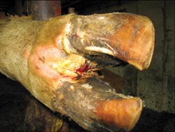
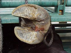
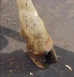
Foot rot is characterized by a sudden onset of lameness. The animals are very painful and will often only touch their toe to the ground. One or more limbs can be affected by foot rot, although it is usually one of the hind limbs. The skin and soft tissue between the toes become red and swollen, which causes the toes to spread apart. A swelling from the top of the hoof to the dewclaws or higher is often observed. Affected cattle will eat less because of decreased grazing time and fewer trips to the feed trough, or they may go off feed altogether. The skin between the toes will develop a crack where protruding dead or decaying tissue will be present. There is a foul odor. If the animal is not treated at this point, the infection may spread to the deeper structures, including bones, tendons, and joints. When this happens, it is considered a chronic condition, and is much harder or impossible to treat. The swelling will become more severe and may go up higher on the leg. Animals will often carry the leg at this point.
A condition named super foot rot has been a problem in some areas. Super foot rot is a strain of F. necroforum that is resistant to multiple antibiotics. Super foot rot will appear the same as regular foot rot in the early stages of the disease, but will progress rapidly and not respond to standard treatments. If a case of foot rot continues to worsen, even after treatment, a veterinarian should be consulted. While foot rot is a major cause of lameness, it should not be assumed that all lameness is due to foot rot. Conditions that can have similar clinical signs are fractures, sole abscesses and abrasions, fescue toxicity, and infected toes. To determine if it is foot rot, the foot must be examined. Only with foot rot will the skin and tissue between the toes be swollen and cracked with decaying tissue protruding.
Foot rot in a calf. There is swelling of the entire foot from above the hoof to above the dewclaws.
Treatment
The most common treatment for foot rot is a long-acting tetracycline (200 mg oxytetracycline) such as LA-200®, Biomycin 200®, Oxycure 200®, and a number of other generic products. The dose is 4.5 cc per 100 pounds of body weight given under the skin (subcutaneously [SQ]). This may be repeated in 48 to 72 hours if needed. Penicillin is sometimes used, but requires daily doses and is an extra label use of this product, so a veterinarian must be consulted before using this. Nuflor® is approved for use in foot rot at 6 cc per 100 pounds of body weight. Nuflor® has been shown to be very effective in the treatment of foot rot (Nuflor Technical Bulletin) but will be more expensive. For this reason, many producers use it for cases that do not respond to initial treatment. Excenel® is also approved in beef cows for foot rot, and only has a three-day meat withdrawal. It would be a good choice if the animal might be marketed soon. Along with antibiotics, flossing between the toes with a clean rope or twine will help remove some of the dead tissue. Keeping the foot clean and dry is helpful. All SQ injections should be given in the neck or in front of the shoulder. If the foot does not improve within two to three days, a veterinarian should be consulted. A veterinarian can remove the infected tissue surgically, or, in severe cases that only involve one toe (or claw), the toe can be amputated. For valuable animals, a claw salvage surgery may be attempted.
Prevention
Prevention is a very important aspect of this disease, and the major focus is to ensure good health of the foot. Rough areas should be smoothed or fenced off from cattle, and debris should be cleared. Sharp gravel should be replaced with smooth stones. To keep cattle from standing in wet, muddy conditions, barnyards should be scraped frequently, and measures to improve drainage should be taken.
Zinc is important in maintaining the integrity of the skin and hoof. In a three-year study, feeding 5.4 gm of zinc methionine per steer daily (added to a free-choice mineral supplement,) decreased the incidence of foot rot, and increased daily gain in grazing steers. Feeding organic iodide (EDDI) at 10 to 15 mg per head per day has been helpful in decreasing the incidence of foot rot on some farms (Maas). Consult with your veterinarian and/or nutritionist when deciding about adding a zinc or organic iodide supplement.
There is a vaccine available that targets the bacteria F. necroforum. This vaccine may be helpful in reducing the incidence and severity of disease, but appears to be the most effective and economical in the feedlot or other intensive rearing situations. Good management programs, including mineral supplementation and vaccination programs increase the overall immunity of the cattle, thereby making them less susceptible to disease.
Summary
Foot rot is a major cause of lameness in cattle and can have a great economic affect on a farm. It is a disease of the skin and other tissues between the toes of all classes of cattle. Swelling and moderate to severe lameness accompanies the condition. Prevention and early treatment, along with good overall management programs, are essential to decrease the incidence and economic effect of the disease.
References
Brazzle F.K. 1993. Cattleman’s Day Report of Progress 704. Agriculture Experiment Station. Kansas State University.
Edmonson, A.J. 1996. Interdigital Necrobacillosis (Footrot) of Cattle. Smith, B.P., ed. Large Animal Internal Medicine 2nd ed., P1314 Mosby, St. Louis, Mo.
Bovine Interdigital Necrosis. In Radostits, O.M. et al. 1994. Veterinary Medicine 8th ed. Baillure Tindal pp. 867-870 London, Philadelphia, Pa.
Nuflor, Technical Bulletin
Maas, John, D.V.M., Extension Veterinarian School of Medicine University of California Davis.


